

The Imaging Division
of the Institut Calot
With a team of around twenty professionals, the Institut Calot's imaging department offers a wide range of examinations.
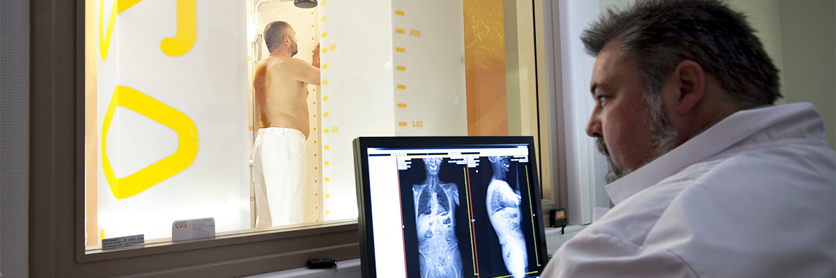
SCANNER AND EOS SYSTEM
Low-dose stereoradiographic images of patients in a functional position.
2D and 3D whole-body measurements in 20 seconds.
Reduced exposure to X-rays :
- 10 times less than traditional radiography
- 100 times less than a CT scan
MRI
Produces detailed images of bone and soft tissue, including the spinal cord and peripheral nerves.
The Institut Calot's MRI is one of the quietest and patient-friendly of its generation, making it much less stressful for the patient.
RADIOLOGy
Traditional radiography is the first and most common medical imaging examination.
X-rays are sent by the machine through the part of the body being studied. They reveal any bone abnormalities
ECHOGRAPHy
Ultrasonography uses ultrasound to analyse tissues. There are no known contraindications to this examination. The ultrasound is produced by a probe placed on your body.
SCINTIGRAPHy
Scintigraphy is a nuclear medicine technique used to analyse bones and organs. Before the examination, a contrast product is injected to highlight the targeted areas.
ECHODOPPLER
A Doppler is associated with ultrasound as a minimally invasive technology. It measures blood flow velocity, flow rate and any vascular resistance.
 USEFUL INFORMATION
How to get here
More information on how to get to our establishments by plane, train, car or ferry...


Hopale in videos